Integrated MIMS Version: March 2016 Login | | Sitemap
Case Study: Cardiac Tamponade

Pericardiocentesis from the sixth intercostal space midaxillary line under transthoracic echo guidance


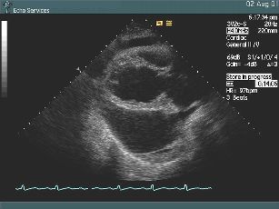
The initial Parasternal Long Axis view gives an understanding to this patient's severe chest pains. The dark anechoic area surrounding the heart is a very large pericardial effusion. The heart swings within this space and there is right ventricle collapse during the cardiac cycle, which are signs of cardiac tamponade. The greatest effusion volume is located posteriorly to the left ventricle.


The left paravertebral view again shows the large posterior effusion. The pericardium is often difficult to observe. In this instance a pleural effusion is also evident. The thin line demarcating the two effusions is then noted as the pericardium.


The appropriate point and angle of entry is critical in assessment prior to pericardiocentesis. The best site of entry presents as the smallest distance to traverse to the effusion and area of largest volume. The 6th intercostal space midaxillary line is chosen and by echo guidance the needle is positioned. A very large pericardial effusion volume of 1700ml is removed initially.


A sufficient time elapses and the draining catheter is removed. The subsequent echo reveals that there is only a trivial residual pericardial effusion. Respiratory collapse has returned to the inferior vena cava and there is no heart swing, or evidence of cardiac tamponade. Posteriorly to the pericardium the pleural effusion has increased in size with the removal of the pericardial volume and pressure.

Copyright © 2008 - 2009 Cardio Vascular Services